Bird Retinas Run Without Oxygen, Upending Vertebrate Metabolism AssumptionsBird Retinas Run Without Oxygen, Upending Vertebrate Metabolism AssumptionsBird Retinas Run Without Oxygen, Upending Vertebrate Metabolism AssumptionsBird Retinas Run Without Oxygen, Upending Vertebrate Metabolism Assumptions
May 14, 2026
A January 2026 study in Nature, led by evolutionary physiologist Christian Damsgaard at Aarhus University, has confirmed that the inner retina of birds operates entirely without oxygen, resolving a...
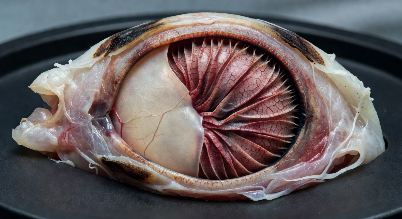
A January 2026 study in Nature, led by evolutionary physiologist Christian Damsgaard at Aarhus University, has confirmed that the inner retina of birds operates entirely without oxygen, resolving a paradox that has puzzled scientists for centuries. The counterintuitive piece is not that birds have exceptional vision, but that the inner retina achieves it using anaerobic glycolysis (the oxygen-free breakdown of glucose), a metabolic pathway roughly 15 times less efficient than the aerobic respiration every other vertebrate retina depends on. The outer retina, which contains the photoreceptor outer segments and retains a sparse vascular supply, still uses oxygen — but the inner retina, where the most demanding neuronal processing happens, does not. For biomedical researchers studying stroke and ischemic injury, the finding offers a concrete biological model of how highly active tissue survives without oxygen.
Retinal tissue is among the most energetically demanding in the animal kingdom, consuming two to three times more energy than the same mass of typical brain tissue. It is built from complex networks of sometimes more than 100 different types of neurons, each demanding a constant biochemical supply. In most vertebrates, including humans, that supply arrives through dense branching networks of blood vessels threaded through the retina. Birds have almost none of that vascular infrastructure. How tissue that hungry could survive without the oxygen that blood vessels deliver had no confirmed answer until the Damsgaard study.
What the Researchers Found
The study's central finding is deceptively simple: bird retinas do not have an unusual oxygen-acquisition adaptation. They survive because they do not use oxygen at all. Damsgaard and his team demonstrated for the first time that avian retinal tissue powers itself entirely through anaerobic glycolysis, the same ancient biochemical pathway that predates atmospheric oxygen by billions of years.
The metabolic arithmetic matters here. In glycolysis, one molecule of glucose is converted into two molecules of pyruvate through a sequence of enzyme-catalyzed reactions. That process releases two molecules of ATP (adenosine triphosphate, the universal energy currency of living cells). Under aerobic conditions, oxygen enables further biochemical reactions that break down pyruvate and produce another 30 molecules of ATP -- making aerobic energy extraction from a single glucose molecule 15 times as efficient.
Bird retinas, by Damsgaard's findings, accept that 15-fold penalty and function anyway. The tissue is, in his words, "one of the most metabolically active tissues in the animal kingdom, yet it worked with no apparent blood perfusion. It was a complete paradox."
The paradox had a structural component that made it so durable. Most vertebrate retinas are richly vascularized because oxygen cannot diffuse far through tissue before being consumed. Birds largely lack retinal blood vessels yet see at extraordinary resolution. The January 2026 Nature paper is the first study to confirm the mechanism resolving that contradiction: the tissue simply does not require what the vessels would deliver.
The Methodology
Damsgaard approached the problem as an evolutionary physiologist, framing the bird retina not as a medical anomaly but as a biological system under selection pressure for millions of years. The question his team posed was whether avascular avian retinas had evolved some hidden oxygen-acquisition mechanism or had done something more radical: eliminated the need for oxygen entirely.
The Aarhus University team measured oxygen supply and metabolic demand in avascular retinal tissue, testing whether the retina showed any evidence of aerobic metabolic activity. The findings ruled out a hidden oxygen pathway. Anaerobic glycolysis accounted for the tissue's energy budget. The pathway converts glucose into pyruvate, and the associated free energy drives ATP synthesis -- all without any requirement for oxygen.
The inner retina appears to run on the two-ATP output from glycolysis alone, relying on glucose throughput rather than oxygen-enabled efficiency. The Damsgaard team's measurements suggest the inner retina demands roughly 2.5 times more glucose than other parts of the bird brain — the metabolic price of running anaerobically at high load.
The pecten oculi (a pleated, heavily vascularized comb-like structure unique to bird eyes, projecting from the optic disc into the vitreous body), reportedly first correctly described in the 17th century, turns out to be the support system that makes this anaerobic strategy viable. The Damsgaard team found that the pecten expresses genes for lactic acid transporters — the proteins that move lactate out of tissue — and identified glucose delivery as its companion role. The pecten is not bringing oxygen INTO the retina, as had been theorized for centuries; it is pumping glucose IN to fuel anaerobic glycolysis and clearing lactate OUT (the metabolic waste of that pathway). That dual function resolves a structure-function question that had been open since the 17th century.
Why It Matters
The energy economics of this finding connect to one of the deepest transitions in the history of life on Earth. Around 3.4 billion years ago, cyanobacteria evolved chlorophyll-based photosynthesis, releasing oxygen as a byproduct of splitting water molecules. The resulting accumulation of atmospheric oxygen -- the Great Oxidation Event (which began, according to geological records, roughly 2.46 billion years ago) -- reoriented the biochemistry of nearly every complex organism that followed. As Damsgaard put it: "We've been hooked on 20% [atmospheric] oxygen for millions of years."
That dependence is both a strength and a vulnerability. In most tissues, oxygen deprivation leads rapidly to cell death. Ischemia (a restriction in blood supply causing a shortage of oxygen and nutrients needed for cellular metabolism) is a primary driver of stroke damage and heart disease mortality across most Western countries. Understanding how any tissue sustains high metabolic activity without oxygen therefore carries direct therapeutic relevance.
The bird retina represents a confirmed, naturally evolved model of that tolerance. The relevance is specific: the retina is not a low-demand tissue tolerating occasional hypoxia; it is one of the most demanding tissues known, running at full load on a biochemical pathway that delivers only 2 ATP per glucose molecule instead of the roughly 30 to 32 ATP that aerobic respiration yields.
- Anaerobic glycolysis yields: 2 ATP per glucose molecule
- Aerobic respiration (glycolysis plus mitochondrial reactions) yields: approximately 30 to 32 ATP per glucose molecule
- Efficiency gap: aerobic metabolism is 15 times more efficient, and sometimes more
- Bird retinal energy demand: 2 to 3 times greater than equivalent brain tissue mass
Competitive Landscape
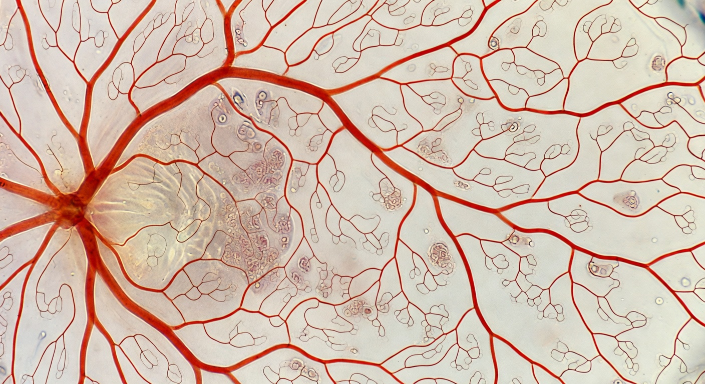
No directly comparable commercial peers were publicly identifiable at publication time in avian retinal biology research. The Damsgaard study is framed in available sources as the first confirmation of the anaerobic mechanism, resolving a paradox that had no prior experimental resolution. Adjacent activity from ischemia and stroke therapeutics research suggests the biomedical application space is active, but a direct competitive ranking of research groups working on this specific finding would require disclosures not yet released.
Bird retinas occupy a confirmed outlier position among vertebrates. Human and most other vertebrate retinas are densely vascularized precisely because oxygen-dependent aerobic metabolism cannot be sustained without local delivery infrastructure. The bird retinal architecture -- lacking those vessels yet supporting networks of sometimes more than 100 different neuron types in tissue that consumes two to three times the energy of equivalent brain mass -- represents one of the more extreme metabolic trade-offs documented in vertebrate sensory systems.
Independent analyst commentary specifically on this announcement was not publicly available at publication time.
Limitations and Caveats
The Damsgaard study resolves the central question of mechanism: bird retinas use anaerobic glycolysis and function without oxygen. What it does not resolve is how the tissue manages the consequences of that choice at scale.
The 15-fold efficiency gap is partially answered by the Damsgaard team's measurements — the inner retina demands roughly 2.5 times more glucose than other parts of the bird brain — but the question of how that glucose flux is sustained at sufficient rates remains open. Differences in neuron packing density or metabolic demand across the retina's more than 100 neuron types are also unresolved. The study does not specify which bird species were examined in detail, making it unclear whether the finding applies uniformly across avian lineages or is concentrated in particular groups.
The Damsgaard study characterizes the pecten oculi's role as dual-purpose — glucose delivery plus lactate clearance via expressed lactic acid transporter genes — but the exact glucose throughput rate, the lactate-handling capacity, and the molecular-level details (which specific transporter isoforms, their expression levels, their regulation) remain to be characterized in follow-up work.
The evolutionary timing now has a window. The Damsgaard team's analysis places the emergence of the oxygen-free inner retina sometime during the dinosaur era — after the avian lineage split from its crocodilian relatives but before modern bird groups diversified. What selection pressure favored losing retinal vasculature — optical clarity from removing vessel shadows, reduced risk of retinal detachment, or something else — remains the open question. The Nature paper bounds the timing; the cause is still under investigation.
What Comes Next
Damsgaard has framed the broader research question in terms that suggest the bird retina is a starting point, not a destination. "What are the extremes of life?" he asked. "How far can we bend the conditions under which highly metabolically active tissues can actually survive?" That framing points toward a comparative physiology program: if one tissue in one vertebrate lineage has evolved full anaerobic function under high metabolic load, what else might be possible, and what structural or biochemical features make it viable?
The most immediate translational interest is in stroke and ischemic injury. Ischemia causes not only oxygen deprivation but also reduced delivery of nutrients and failure to clear metabolic waste. Any tissue that can maintain function under those conditions is a biological proof-of-concept for therapeutic intervention. If the mechanisms that allow bird retinas to run anaerobically can be identified at the molecular level, they may point toward strategies for protecting human brain or cardiac tissue during the critical window between ischemic event onset and clinical intervention.
The pecten oculi remains a target for follow-up biochemistry. Damsgaard's team established the dual glucose-in / lactate-out function, but the molecular-level questions — which specific lactic acid transporter genes are expressed, how glucose delivery scales with retinal demand, how the pecten maintains pH balance under high lactate efflux — could substantially refine the picture. Comparative studies across avian lineages (raptors with their extraordinary visual acuity, nocturnal birds with different photic loads, flightless species with smaller eyes) are an obvious next step in mapping the limits of the strategy.
For neuroscientists and biomedical engineers designing retinal implants or ex vivo tissue models, this finding shifts a baseline assumption that has constrained scaffold architecture for decades: vascularization of retinal constructs is not a biological requirement, it is a metabolic workaround. A retina running on 2 ATP per glucose and no oxygen supply is not a broken system -- it is a design principle. Any scaffold or organoid that can guarantee sufficient glucose delivery without embedding a vascular network just gained a confirmed natural precedent, one that has been sustaining more than 100 neuron types at two to three times brain-tissue energy demand for millions of years.
-- Zara Velez, Emerging Technology Editor
Sources: Quanta Magazine -- How the Bird Eye Was Pushed to an Evolutionary Extreme - Damsgaard et al., Nature, January 2026 (Aarhus University)
More on Revuzia
Suniva Plans 564-Job Solar Manufacturing Facility in South Carolina
Gene Therapy ETX101 Cuts Dravet Seizures 76% — and Tracks Toward Neurotypical Development
Algorithmiq Raises EUR 18 Million, Beats Harvard and Stanford on Quantum Drug Simulation